A knee MRI scan is one of the most useful diagnostic tools available to an orthopaedic surgeon. It does not involve radiation, it images soft tissue that X-rays cannot, and it often provides the single most complete picture of what is going on inside your knee.
This article explains when a knee MRI is needed, what a knee MRI shows, how it performs in diagnosing injuries like ACL tears, and what knee MRI costs in Mumbai look like in practice. Whether your doctor has recommended one or you are trying to understand your options before a consultation, the answers are here.
What Is a Knee MRI Scan?
MRI stands for magnetic resonance imaging. A knee MRI scan uses a strong magnetic field and radio waves to produce detailed cross-sectional images of the knee joint. Unlike X-rays, it produces no ionising radiation. Unlike ultrasound, it can image deeper structures such as cartilage, ligaments, and the bone beneath the surface.
The scan typically captures multiple planes of the knee, including front-to-back, side-to-side, and cross-sectional slices. This gives the radiologist and orthopaedic surgeon a three-dimensional understanding of the joint without making a single incision.
Two main types of MRI machines are used:
- 1.5 Tesla MRI: The standard option used in most hospitals and diagnostic centres. Reliable for the vast majority of knee diagnoses.
- 3 Tesla MRI: A higher-field-strength machine offering greater image resolution. Useful in complex or ambiguous cases. A prospective study comparing both noted that routine use of 3.0T MRI did not significantly improve accuracy over 1.5T for diagnosing ACL and meniscal injuries in most clinical situations.
Contrast dye is occasionally used in knee MRI scans, most often when the surgeon suspects a synovial abnormality, infection, or tumour. In most straightforward cases, contrast is not required.
When Is a Knee MRI Needed?
Not every knee problem requires an MRI. Your doctor will decide based on your symptoms, clinical examination findings, and what information is needed to plan treatment.
A knee MRI is typically recommended when:
- There is suspected ligament damage, particularly an ACL tear or MCL injury
- A meniscus tear is suspected following a twisting injury or gradual onset of knee clicking and locking
- Knee pain persists beyond 4 to 6 weeks without a clear cause on clinical examination
- There is significant swelling that appeared rapidly after an injury
- An MPFL injury or patellar instability is being assessed
- Cartilage damage or early knee osteoarthritis needs grading prior to considering surgery
- A popliteal cyst (Baker’s cyst) or other soft tissue mass around the knee needs characterisation
- Post-operative assessment is required after ligament reconstruction or meniscus repair
- There is unexplained bone pain, and a stress fracture or bone marrow lesion is suspected
A knee MRI scan is not always the first investigation. An X-ray is usually done first to assess bone alignment and rule out fracture. The MRI follows when soft tissue information is needed.
Not every positive clinical finding requires MRI confirmation before treatment. A clearly positive Lachman test in the context of a known twisting injury is often sufficient to plan ACL reconstruction without waiting for imaging.
What Does a Knee MRI Show?
A knee MRI scan produces images of virtually every structure inside and around the knee joint. This is what makes it so clinically useful.
Ligaments
The ACL (anterior cruciate ligament), PCL (posterior cruciate ligament), MCL (medial collateral ligament), and LCL (lateral collateral ligament) are all visible on MRI. A complete tear appears as a discontinuity in the ligament fibres. Partial tears, swelling, and chronic scarring also have distinct appearances.
Menisci
The medial and lateral menisci, the two cartilage discs that cushion the knee, show up clearly. Tears appear as bright lines or signals within the meniscal tissue on specific MRI sequences. MRI has been shown to have high sensitivity for medial meniscal tears, typically above 88 to 91%, according to multiple studies including research published in the Khyber Medical University Journal (2023).
Articular Cartilage
The cartilage covering the femoral condyles, tibial plateau, and patella is visible. MRI can identify thinning, softening, fissuring, and full-thickness loss. It is less sensitive for very early cartilage changes than for ligament injuries.
Bone
Bone marrow oedema, stress fractures, and subchondral bone lesions appear as bright areas on specific sequences. These are often missed entirely on plain X-rays.
Tendons
The quadriceps tendon, patellar tendon, iliotibial band, and biceps femoris tendon are all evaluable. Tendon thickening, tearing, or calcification can be identified.
Bursae and Joint Fluid
Excess joint fluid, synovial thickening, bursitis, and soft tissue masses such as popliteal cysts are clearly seen on MRI.
What a Knee MRI Cannot Show
MRI has limitations. It does not reliably diagnose every cartilage lesion. A 2023 study in the Journal of Experimental Orthopaedics specifically found that diagnostic sensitivity for cartilage injuries was significantly lower than for ACL and meniscal injuries. For definitive cartilage assessment, knee arthroscopy remains the most accurate method.
Knee MRI for ACL Tear
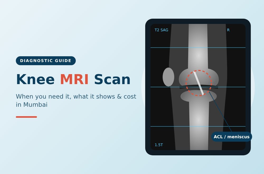
Knee MRI for ACL tear is one of the most common uses of this imaging modality. A torn ACL typically appears on MRI as a discontinuity, increased signal, or abnormal angulation of the ligament on T2-weighted sequences. Associated bone bruising on the lateral femoral condyle and posterolateral tibial plateau is also a common finding that reinforces the diagnosis.
Research published in the Annals of Pakistan Institute of Medical Sciences (2023) found that MRI had a sensitivity of 89.9% and an accuracy of 88.6% for diagnosing ACL injuries. A separate study published in the Khyber Medical University Journal (2023) reported sensitivity of 93.4% for ACL injuries with high negative predictive value, meaning that a normal MRI makes an ACL tear unlikely.
MRI is also valuable in the context of ACL tears because it allows the surgeon to assess for associated injuries simultaneously. Meniscal tears accompany ACL injuries in around 30% of cases. Bone bruises and cartilage damage, which affect surgical planning and recovery expectations, are also identified on the same scan.
For patients planning ACL reconstruction surgery, the pre-operative MRI helps the surgeon understand the degree of ligament disruption, identify any partial tears, and assess the posterior cruciate ligament and overall joint condition before deciding on surgical approach.
That said, MRI does not replace clinical examination. The Lachman and pivot shift tests remain essential. In experienced hands, clinical assessment combined with MRI findings provides the most reliable basis for surgical decision-making.
Knee MRI Cost in Mumbai
Knee MRI cost in Mumbai varies depending on where the scan is done, the strength of the machine, and whether contrast is used. Mumbai, as a Tier 1 city, generally sits at the upper end of the national price range for diagnostic imaging.
Indicative Price Range
| Type | Approximate Cost (INR) |
|---|---|
| Standard knee MRI scan (1.5T, no contrast) | ₹4,500 to ₹8,000 |
| Advanced knee MRI scan (3T, no contrast) | ₹6,000 to ₹12,000 |
| Knee MRI with contrast | ₹8,000 to ₹15,000 |
These are indicative ranges based on publicly available pricing data. The Manipal Cigna cost guide cites an average MRI knee scan cost in Mumbai of around ₹7,500, though actual costs at individual centres vary. Prices at government hospitals or diagnostic centres with CGHS or NABH empanelment are typically lower than private hospital rates.
Factors That Affect the MRI Knee Scan Cost
- Facility type: Private hospitals generally charge more than standalone diagnostic centres.
- Machine strength: 3 Tesla machines cost more to operate and scan on.
- Contrast use: Contrast-enhanced scans involve additional material and monitoring cost.
- Radiologist fees: The cost of the written radiological report may or may not be included in the quoted scan price.
- Insurance: Many health insurance policies cover knee MRI scans if ordered by a registered medical practitioner with a clinical indication. Check with your insurer whether pre-authorisation is required.
Costs vary between hospitals and individual cases. Always obtain a specific price quote before scheduling, and confirm what is included in the price, such as the scan, report, and any radiologist consultation.
What to Expect During a Knee MRI Scan
A knee MRI scan is painless and safe for most people. Knowing what to expect can reduce any anxiety about the process.
Before the Scan
You will be asked to remove any metal objects, including belts, jewellery, hair clips, and watches. If you have metal implants such as a pacemaker, cochlear implant, or certain types of surgical clips, tell your radiologist before the scan, as some metal devices are incompatible with the MRI magnetic field. Most modern orthopaedic implants, including knee replacement implants, are MRI-compatible, but always confirm.
During the Scan
You lie on a padded table with your knee positioned inside or near the MRI coil. The table slides into the scanner. A knee MRI typically takes between 30 and 45 minutes. During the scan, you will hear loud rhythmic knocking sounds; these are normal and indicate the machine is working. You will be given earplugs or headphones. The radiographer can hear and speak to you throughout.
After the Scan
There is no recovery period. You can drive, eat, and continue normal activity immediately after. If contrast was used, a radiographer will monitor you briefly before you leave. The radiologist’s report is typically available within 24 to 48 hours, and your orthopaedic surgeon will review the images alongside the report.
Is a Knee MRI Uncomfortable for People with Claustrophobia?
The knee is scanned in an open or peripheral coil in many machines, meaning your head and most of your body remain outside the scanner. This makes knee MRI more comfortable than brain or spine MRI for those who feel anxious in enclosed spaces.
Research Insight: Dr Amyn Rajani on MRI and ACL Diagnosis
Dr Amyn Rajani is a Consultant Knee, Shoulder, and Hip Surgeon with over 25 years of experience. He is affiliated with Breach Candy Hospital, Saifee Hospital, and PD Hinduja Hospital in Mumbai, and is a reviewer for the Journal of Arthroplasty and the Indian Journal of Orthopaedics.
One of Dr Rajani’s published studies has a direct bearing on how knee MRI findings relate to surgical decision-making. In a paper titled “Triangular notch morphology in young males linked to increased risk of non-contact anterior cruciate ligament tears”, published in the Journal of Arthroscopic Surgery and Sports Medicine (February 2025), Dr Rajani and his co-authors investigated whether the shape of the intercondylar notch on MRI could predict non-contact ACL tears in young males.
This is clinically relevant for the MRI context because it illustrates how a knee MRI scan provides information beyond simply confirming or ruling out a current injury. Notch morphology, which is visible on MRI, may be a risk factor for future ACL tears. The findings suggest that pre-season or screening MRI in young athletes could offer prognostic information beyond just injury detection.
Dr Rajani’s additional published work on ACL reconstruction outcomes further contextualises the importance of accurate pre-operative imaging in planning surgical approach and predicting recovery. His full publications list is available for patients who wish to review the research base that informs his clinical practice.
FAQs about Knee MRI Scan
What does a knee MRI show?
A knee MRI scan shows ligaments, menisci, cartilage, tendons, bone marrow, and surrounding soft tissue. It can identify tears, inflammation, bone bruising, cysts, and signs of arthritis that do not appear on a standard X-ray.
When is a knee MRI needed?
A knee MRI is typically recommended after a significant knee injury, when soft tissue damage is suspected, or when knee pain persists without a clear diagnosis after clinical examination and X-ray. Your orthopaedic surgeon will advise based on your specific presentation.
What is the knee MRI cost in Mumbai?
Knee MRI scan price in Mumbai generally ranges from approximately ₹4,500 to ₹12,000, depending on the facility and the type of machine. Contrast-enhanced scans cost more. Always obtain a specific quote and confirm what the price includes.
How accurate is a knee MRI for ACL tears?
Knee MRI for ACL tear has been reported with sensitivity between 89% and 93% across multiple studies. It is a reliable screening tool, though clinical examination adds important context that imaging alone cannot always capture.
Is a knee MRI safe?
Yes. MRI uses no ionising radiation and is considered safe for the vast majority of patients. Exceptions include those with certain pacemakers, cochlear implants, or specific metal implants. Always inform the radiology team about any implanted devices before the scan.
Does health insurance cover knee MRI cost in Mumbai?
Most health insurance policies cover knee MRI scan cost when ordered by a registered doctor with a documented clinical reason. Check with your insurer whether the scan requires pre-authorisation or a specific referral before you book.